
An MRI-guided targeted biopsy is significantly better than fusion-guided biopsy, says a new study published in March 2021.
A revolution is transforming how doctors diagnose prostate cancer. Traditionally, they have relied on a 12- to 14-needle biopsy guided by transrectal ultrasound (TRUS). However, this method often proves unreliable because it cannot distinguish cancerous tissue. Doctors call it a ‘systematic’ biopsy because they divide the prostate into six sections on each side and take at least one tissue sample from each section, totaling a minimum of 12 samples.
- It often detects insignificant prostate cancers that are unlikely to cause harm or require treatment.
- It frequently misses or fails to detect significant prostate cancers.
- It carries potential side effects, including infection, and the risk of underestimating more aggressive cancers, which could lead to recurrence after surgery or radiation.
The new, revolutionary diagnostic approach is to target only a few needles into the most suspicious areas of the prostate. This method significantly increases the chances of capturing the oldest, most aggressive cancer cells—those that are located where the tumor first developed—while also reducing the risk of infection and other side effects.
The key to this targeted approach is multiparametric MRI (mpMRI). Before the biopsy, an mpMRI scan is excellent at detecting significant prostate cancer. A skilled radiologist assigns a PI-RADS score from 1 to 5 based on MRI results. A score of 5 signals the highest likelihood of prostate cancer. Doctors see a score of 4 or 5 as a strong sign of significant cancer. They use this score to guide where to take biopsy samples.
Lab specialists examine biopsy samples and assign a Grade Group score from 1 to 5. A Grade Group of 2 or higher confirms significant prostate cancer.
What is the best method for targeting? A recent study by Prince et al. (2021)[i] compared real-time in-bore MRI-guided biopsy with fusion-guided biopsy.
“In-bore” refers to the process where the radiologist is able to see the tumor’s size, shape, and location in real-time while the patient is inside the MRI machine. This is made possible by the 3D, high-resolution capabilities of the MRI.
This method provides precise control and allows the radiologist to confirm the exact placement of a minimal number of needles (usually 2-4) directly into the core of the suspicious area. As a result, this biopsy technique has the smallest margin for error.
Fusion guidance uses MRI and ultrasound images aligned through digitized co-registration. It matches prior MRI scans with real-time ultrasound images during the procedure. This process forces a fit between MRI scans from a patient lying on their back and ultrasound images from a patient lying on their side. Urologists invest in fusion guidance to improve patient care. They believe MRI is superior to TRUS for targeting prostate tumors. Fusion guidance is seen as a way to bring MRI advantages into the ultrasound-guided procedure.
The Prince study highlights an issue with fusion guidance. It shows fusion can cause misregistration error due to prostate deformation. This deformation occurs between MRI scans and real-time ultrasound views. The fusion process does not account for these changes in the prostate’s shape.
As a result, the needle might miss the actual tumor. Even small errors can have major consequences for the patient. For example, an aggressive cancer like GG3 could go undetected. This increases the risk of recurrence and worsens patient outcomes.
Some urology practices may refer to their fusion-guided procedures as “MRI-guided targeted biopsies,” but this is misleading. Moreover, to address the targeting errors inherent in fusion, many of these biopsies also include a standard 12-needle systematic biopsy, in addition to the 2-4 targeted needles indicated by the software!
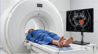
Technicians perform MRI imaging in real time while the patient lies inside the MRI machine’s bore (tunnel).
At our Center, we position the patient on their back inside the magnet and choose not to use an endorectal coil (inserted into the rectum).
Real-time MRI imaging remains free from inaccuracies or distortion across multiple imaging sequences.
MRI sequences distinguish tumor tissue from normal prostate tissue, allowing the radiologist to precisely identify the size, shape, and location of the targeted tumor in real time.
Our prostate radiologists are highly experienced in interpreting MRI scans.
MRI-guided biopsy typically requires 2-4 needle samples.
This method eliminates the extra time spent on co-registration, which would otherwise involve gland segmentation and point-by-point alignment.
Transrectal ultrasound is performed in real time in the urology exam room, but previously captured MRI scans are not live when integrated into the fusion software.
The patient lies on their side on the examining table with the ultrasound probe inserted rectally. This different positioning introduces a margin of error during the point-by-point co-registration (fusion) of the prostate outline.
Fusion images may have some distortion, however small, due to
– Co-registration error
– Patient movement
– Patient breathing, coughing or sneezing
The urologist views the target based on images from a previous MRI fused with real-time ultrasound. However, real-time ultrasound cannot differentiate between tumor and normal prostate tissue within the gland, causing the software to create an artificial model of the prostate, which depicts the size, shape, and location of the targeted tumor.
Urologists must learn to read MRI images.
Fusion targeting typically involves 2-4 needles directed into the computer-generated target, but fusion-guided biopsies often include an additional 12-needle systematic biopsy to compensate for potential inaccuracies in the fusion process.
The procedure requires extra time for point-by-point co-registration and image matching.
MRI targeting performed in the bore (tunnel) of the magnet surpasses fusion, which is considered a less accurate substitute for the precision of real-time MRI. The Prince study highlights:
The precision of these two techniques is especially important if target-only diagnosis becomes the new standard. Both the PROMIS and PRECISION trials support this change in approach. They found that using only MRI-targeted biopsies improves the accuracy of prostate cancer diagnosis. This method avoids the need for traditional systematic biopsies and leads to more reliable results.
The authors compared the target-specific cancer detection rates of in-bore MRI versus fusion using a study sample of 286 men with mpMRI PI-RADS categories of 4 or 5.
The findings of the Prince study revealed that in-bore MRI-targeted biopsy of the prostate has a significantly higher target-specific cancer detection rate than fusion MRI-targeted biopsy.