

Doctors have used TRUS biopsy to detect prostate cancer for the past 20 years. They call it a “blind” and random process because it cannot specifically identify certain tumors, especially very small ones MRI-Guided Biopsy .
Ultrasound imaging cannot pinpoint precise cancerous areas, so it fails to detect many tumors. TRUS biopsies typically target the outer regions of the prostate but often miss 30-40% of cancer in the anterior, midline transition zone, or apex—this problem is called sampling error. To compensate, doctors take 12-14 needle samples during TRUS biopsies, and sometimes “saturation” biopsies use up to 24 needles. However, increasing needle numbers raises the risk of discomfort, infection, and side effects like urinary or sexual issues, yet they still fail to identify all tumors. No other cancer is biopsied this way.
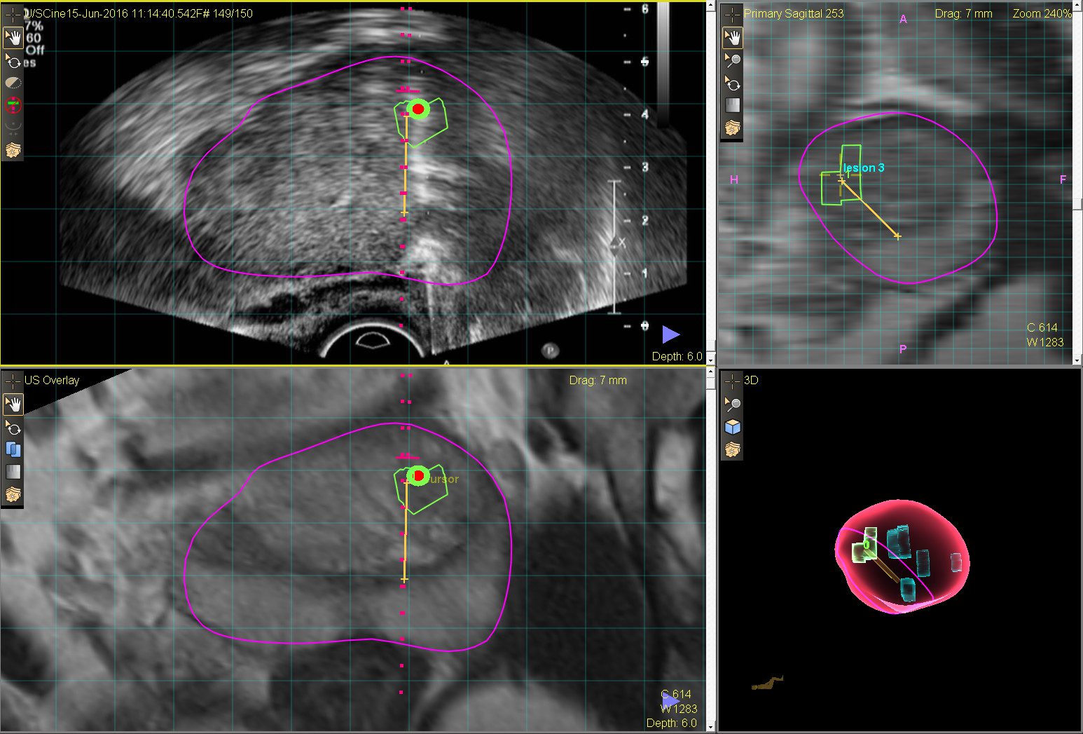
Our BlueLaser™ 3T mpMRI provides high-resolution, 3D images that clearly differentiate healthy from diseased tissue, allowing for precise localization of suspicious tumors. This advanced imaging technique offers a superior method for characterizing and targeting malignancies, particularly important when diagnosing smaller, lower-risk tumors.
An MRI-Guided Biopsy precisely targets the tissue identified in the MRI scan, ensuring that tissue samples are taken from the areas most likely to contain cancer. This targeted approach contrasts with the “blind” technique of a TRUS biopsy, offering greater confidence for both physicians and patients in the accuracy of the diagnosis.
Biopsy samples are analyzed in top-tier laboratories by expert pathologists. Depending on the patient’s individual risk factors, further analysis can include evaluating molecular markers and testing gene sequences for harmful mutations. If necessary, genomic testing is used for the most comprehensive results. These findings, when correlated with MRI images, provide a definitive confirmation of prostate cancer. If a tumor is identified as low- to moderate-risk, it may qualify for focal treatment.
Some doctors use fusion-guided biopsy and call it MRI-guided biopsy, but this term misleads patients. Fusion combines previously captured MRI images with real-time ultrasound using software. This method reduces accuracy because patient positioning or slight movement during ultrasound can differ from the original MRI scan. These discrepancies cause fusion-guided biopsies to be less accurate than real-time MRI guidance performed inside the MRI scanner (in-bore). Although fusion improves on TRUS alone, it remains less precise than real-time MRI-guided biopsies.
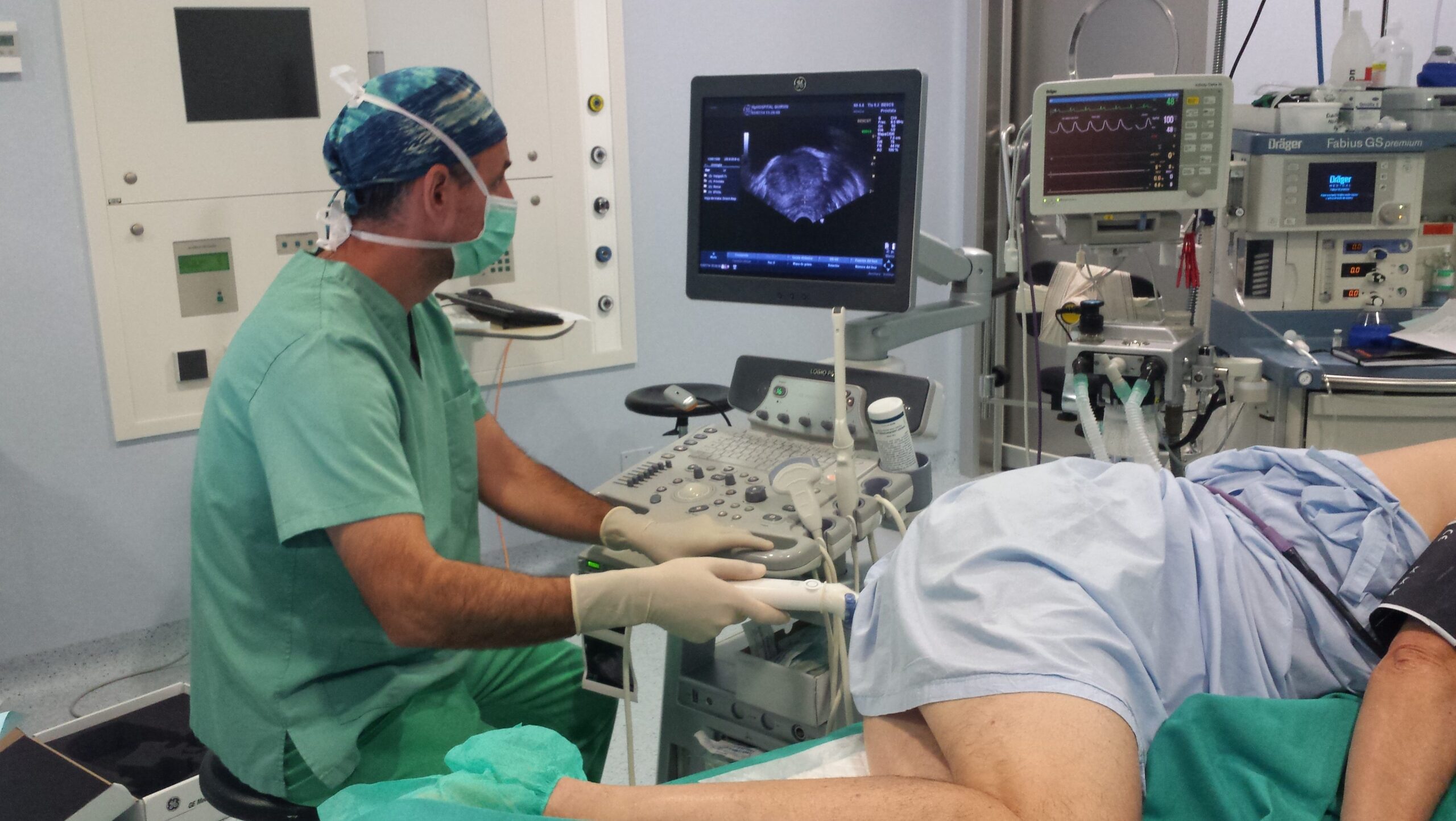
Doctors rely on needle biopsy as the only reliable method to diagnose suspected prostate cancer. They insert thin, hollow needles into the prostate to gather tissue samples. Pathologists then analyze these samples under a microscope. This examination determines whether the cells are cancerous, the tumor size, and the cancer’s aggressiveness. Additionally, doctors can use these samples for genomic or molecular testing to assess how dangerous the cancerous cells are.
We suggest you schedule a BlueLaser™ 3T mpMRI scan of the prostate. PSA levels do not exclusively indicate prostate cancer; noncancerous conditions like inflammation, infection, or BPH can also raise them. An MRI scan provides the best next step to confirm or rule out prostate cancer. If you need a biopsy, the BlueLaser™ 3T mpMRI-Guided Biopsy offers a less invasive and more precise option than traditional TRUS or fusion-guided biopsies.
Ultrasound cannot distinguish between healthy and cancerous tissue, so standard TRUS biopsies miss accurate diagnoses in at least 30% of cases. Biopsy samples often fail to provide complete or accurate results. At our Center, we perform BlueLaser™ 3T mpMRI-Guided Biopsy using fewer needles to precisely target the tumor’s core. You can trust our method to deliver the most accurate results, especially if you consider Active Surveillance or Focal Laser Ablation (FLA).
Studies show that today’s 3T multiparametric MRI detects prostate cancer tumors. Our BlueLaser™ 3T mpMRI provides detailed images of suspicious areas, and certain imaging sequences help assess cancer aggressiveness. However, only laboratory analysis of biopsy samples reveals cellular and molecular details about the tumor.